Home » Without Label » Bones In Leg Diagram / Femur Definition Function Diagram Facts Britannica : Transferred from en.wikipedia to commons by rocket000 using commonshelper.
Bones In Leg Diagram / Femur Definition Function Diagram Facts Britannica : Transferred from en.wikipedia to commons by rocket000 using commonshelper.
Bones In Leg Diagram / Femur Definition Function Diagram Facts Britannica : Transferred from en.wikipedia to commons by rocket000 using commonshelper.. The foot bones shown in this diagram are the talus, navicular, cuneiform, cuboid, metatarsals and calcaneus. The medial, larger bone of the lower leg. He leg's main function in the human is for locomotion and support of the rest of the body. This page is about leg bones diagram,contains aluminium plant safety: The human leg consists of 8 bones, 4 per leg.
He leg's main function in the human is for locomotion and support of the rest of the body. The blood supply to and/or from the navicular bone is disrupted. The ankle of human body. The patella (kneecap) is the sesamoid bone in front of the knee. The tibia and the fibula, at the top of the ankle joint.
Muscular Function And Anatomy Of The Lower Leg And Foot Video Lesson Transcript Study Com from study.com The femur, or thighbone, is the longest and largest bone in the human body. The proximal portion of the tibia is tibial plateau which acts as a cusp for the knee, the distal portion tapers into the medial malleoli and the concave surface which articulates with the talus at the ankle joint. There are in all 7 bones, which fall under tarsal bones category. Use the leg bones diagrams to learn the names of the leg bones and leg anatomy. Here is a horse skeleton with the bones of its front leg colored in a specific way. Wa state leap mittee leap is a bipartisan. Bone diagram forehead (frontal bone) nose bones (nasals) cheek bone (zygoma) upper jaw (maxilla) lower jaw (mandible) breast bone (sternum) upper arm bone (humerus) lower arm bone (ulna) thigh bone (femur) collar bone (clavicle) toe bones (phalanges) ankle bones (tarsals) kneecap (patella) shin bone The femur is the largest bone in the body and the only bone of the thigh (femoral) region.
The femur is the single bone of the thigh.
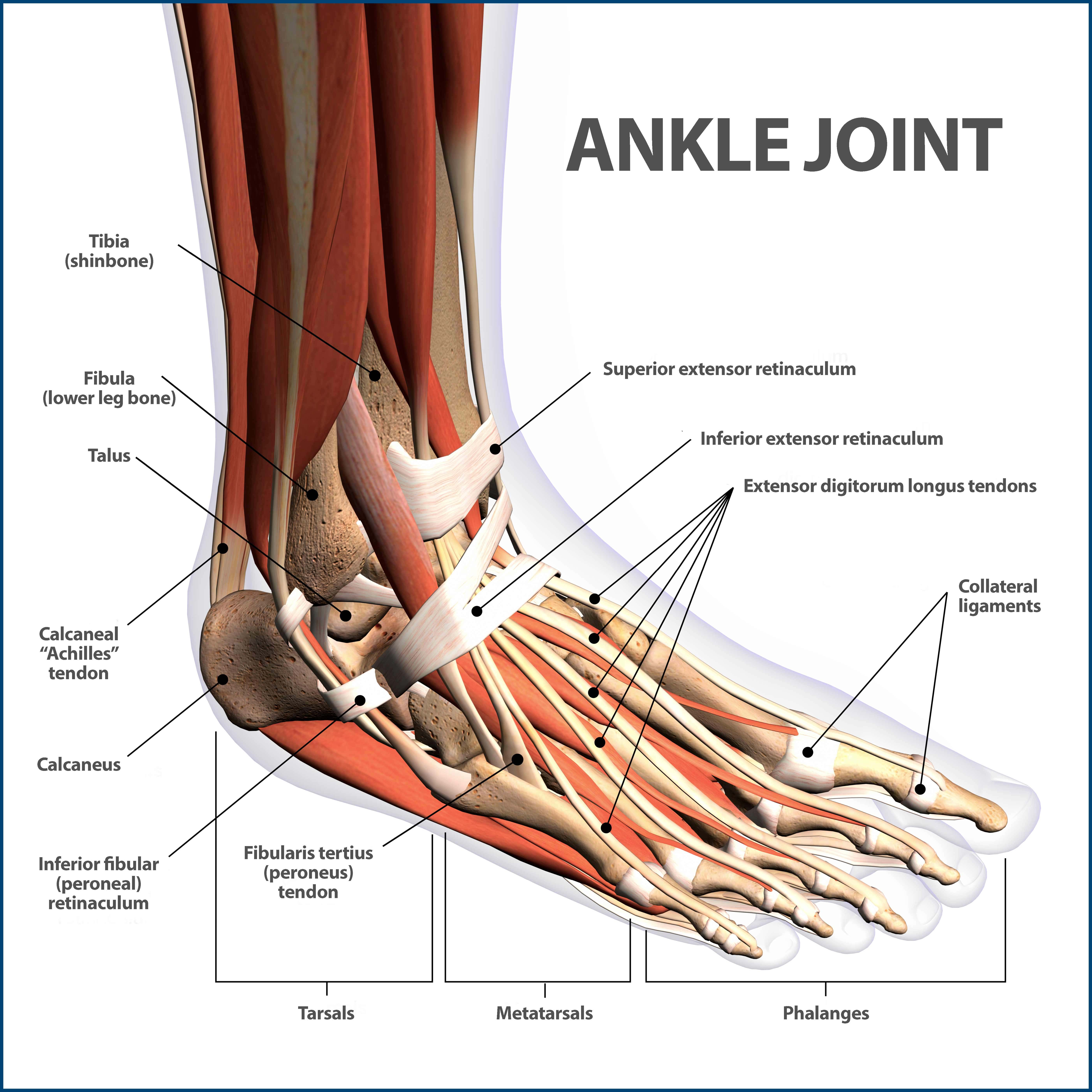
Bone of pelvis pics 12 photos of the bone of pelvis pics , bone. The human leg, in the general word sense, is the entire lower limb of the human body, including the foot, thigh and even the hip or gluteal region. Bone on side of the foot To explain the term in layman's language, it is the heel bone in the skeletal system. As these muscles contract and relax, they move skeletal bones to create movement of the. Its lower end helps create the knee joint. Bone on side of the foot. The medial, larger bone of the lower leg. This area is commonly referred to as the calf. Master leg and knee anatomy using our. The lower leg contains two major long bones, the tibia and the fibula, which are both very strong skeletal structures. He leg's main function in the human is for locomotion and support of the rest of the body. The diagram of bones in the ankle and foot is given below:
Electrical wiring diagrams leg bones diagram femur which are in coloration have a bonus above when looking at any leg bones diagram femur wiring diagram, get started by familiarizing your self. Inflammation of navicular bone and/or bursa. The lower leg is comprised of two bones, the tibia and the smaller fibula. The tibia, commonly known as the 'shin bone', is the largest and most medial of the two.you can palpate its anterior border when you run your finger down the anterior aspect of your leg. The femur, or thighbone, is the longest and largest bone in the human body.
Ankle Fractures Broken Ankle Florida Orthopaedic Institute from www.floridaortho.com The femur is the largest bone in the body and the only bone of the thigh (femoral) region. The patella is the kneecap and articulates with the distal femur. It is likely that abnormal biomechanical stresses are the basis for the disease. This area is commonly referred to as the calf. Also called the shin bone, the tibia is the longer of the two bones in the. Connecting the pelvic girdle to the lower leg is a bone in the thigh area called the femur. He leg's main function in the human is for locomotion and support of the rest of the body. The blood supply to and/or from the navicular bone is disrupted.
The bones together make up the hip.
At the same time, the bones and joints of the leg and foot must be strong enough to support the body's weight while remaining. Leg pain can also be caused by blood clots, varicose veins or poor circulation. These bones provide a groove to hold the tendons. As these muscles contract and relax, they move skeletal bones to create movement of the. Muscles the majority of muscles in the leg are considered long muscles, in that they stretch great distances. The medial, larger bone of the lower leg. The femur is the single bone of the thigh. The bones together make up the hip. Also called the shin bone, the tibia is the longer of the two bones in the. The patella is the kneecap and articulates with the distal femur. Inflammation of navicular bone and/or bursa. Its lower end helps create the knee joint. Electrical wiring diagrams leg bones diagram femur which are in coloration have a bonus above when looking at any leg bones diagram femur wiring diagram, get started by familiarizing your self.
The human leg, in the general word sense, is the entire lower limb of the human body, including the foot, thigh and even the hip or gluteal region. This image is an edited version of this image that was created by user:ladyofhats (mariana ruiz villarreal). Its lower end helps create the knee joint. Also called the shin bone, the tibia is the longer of the two bones in the. Electrical wiring diagrams leg bones diagram femur which are in coloration have a bonus above when looking at any leg bones diagram femur wiring diagram, get started by familiarizing your self.
The Leg Ankle And Foot Amboss from media-us.amboss.com The thigh bone, or femur, is the large upper leg bone that connects the lower leg bones (knee joint) to the pelvic bone (hip joint). The lower leg is comprised of two bones, the tibia and the smaller fibula. Connecting the pelvic girdle to the lower leg is a bone in the thigh area called the femur. The proximal portion of the tibia is tibial plateau which acts as a cusp for the knee, the distal portion tapers into the medial malleoli and the concave surface which articulates with the talus at the ankle joint. Most of the leg skeleton has bony prominences and margins that can be palpated and some serve as anatomical landmarks that define the extent of the leg. Health diagram bone skeleton leg knee science anchor chart human human body. The femur is the single bone of the thigh. Leg bones labeled (page 1).
Bone on side of the foot
10 october 2007 (original upload date) source: The bones of the leg are the femur, tibia, fibula and patella. Bone diagram forehead (frontal bone) nose bones (nasals) cheek bone (zygoma) upper jaw (maxilla) lower jaw (mandible) breast bone (sternum) upper arm bone (humerus) lower arm bone (ulna) thigh bone (femur) collar bone (clavicle) toe bones (phalanges) ankle bones (tarsals) kneecap (patella) shin bone On either side of the cannon bone are the splints that help support the carpus bones of the knee. He leg's main function in the human is for locomotion and support of the rest of the body. The medial, larger bone of the lower leg. Some types of leg pain can be traced to problems in your lower spine. The bones of the leg and foot form part of the appendicular skeleton that supports the many muscles of the lower limbs. The knee joint is the largest joint in the body and is primarily a hinge joint, although some sliding and rotation occur. These muscles work together to produce movements such as standing, walking, running, and jumping. Master leg and knee anatomy using our. Transferred from en.wikipedia to commons by rocket000 using commonshelper. Muscles the majority of muscles in the leg are considered long muscles, in that they stretch great distances.